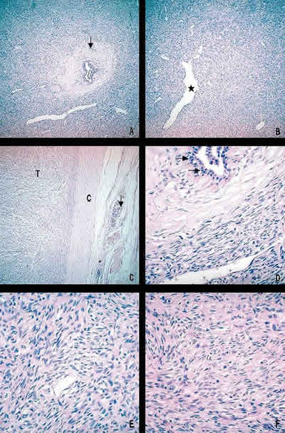
Fig. 2: Microscopic finding. A. Typical histopathologic aspect of ESFT showing hypocellular (peri.ductular, [arrow]) and hypercellular areas (H-E, 100X). B. Characteristic branching haemangiopericytoma-like vessels (asterisk) (H-E, 100X). C. Well demarcated, encapsulated (C) tumour (T), normal breast tissue is seen (arrows) (H-E, 100X). D. Detail of peri-ductular tissue, showing hypocellular area and normal secretory epithelium (arrows) (H-E, 400X). E,F. Characteristic spindle cell tumour cells with indistinct borders and disperse chromatin within vesicular nuclei. No cytologic atypia or necrosis was seen (H-E, 400X).